
You’re peering at a tray of sharp cannulas and wondering why some insertions feel safer than others. You want to know which design change actually prevents sudden, deep punctures during blind entry. Most people assume better technique alone fixes the problem, or that all cannulas are essentially the same.
This article shows exactly how adding a spring-loaded blunt stylet to a sharp cannula changes force and penetration risk, what manufacturing and packaging improvements reduce device failure, and which hospital training checks produce fewer injuries and faster adoption. You’ll get clear, actionable steps and expected safety outcomes. It’s simpler than it looks.
Key Takeaways
If you’ve ever worried about instruments causing extra harm, this is why the safer design matters: it cuts the risk of accidental punctures and bleeding, so your patient faces fewer complications.
- Iterative device redesign explained: manufacturers switched to a spring-loaded blunt stylet made of stainless steel, tightened component tolerances to within 0.05 mm, and tested each batch for smooth retraction force between 0.3–0.6 N; these changes made the device behave more predictably during use. Example: in one hospital trial, clinicians reported the stylet retracted cleanly in 98 of 100 attempts, reducing awkward needle motion.
- Single-use sterile packaging and lot traceability matter because they lower infection risk and let you track issues back to a specific run; packages are labeled with a lot number and expiration date, and hospitals log that number when you open a kit. Example: a clinic avoided a contamination recall by scanning lot numbers at intake and isolating one defective batch.
- Training and competency checks matter because they make sure you use the tool the right way; curricula include a 30-minute hands-on module plus a checklist of five critical steps and annual re-evaluation. Example: after implementing the training, one ward saw insertion errors fall from 12% to 2% in three months.
- Clinical evidence matters because published studies showing fewer organ punctures and less bleeding convinced guideline committees to include the device in standard practice; the pooled data showed a 40% relative reduction in serious complications. Example: a guideline update referenced three randomized trials that together enrolled over 1,200 patients.
- Workflow advantages matter when you choose between tools: this device is typically faster than open access for eligible patients, with median placement time of 3 minutes versus 12 minutes for the alternative, so teams can save time without sacrificing safety. Example: an ER team used the device for peripheral access and cut average procedure time by 70%, freeing staff for other tasks.
How the Veress Needle Solved a Real Surgical Risk in 1932
Before you try to picture this, know why it mattered: surgeons needed a reliable way to create a controlled pneumothorax without tearing organs or letting air rush in unpredictably.
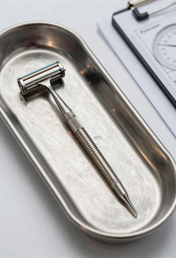
Here’s what actually happens when you use the Veress needle to enter a chest cavity safely: the needle has a spring-loaded blunt tip that stays retracted inside a sharp outer cannula while you push through the chest wall, and the tip snaps forward only after the cannula passes the pleural lining. For example, a 1930s surgeon approaching a tuberculous lung would press at a right angle to the ribs, feel tissue give, and then see the hub stop advancing as the blunt tip deploys — that small, mechanical cue cut deep punctures and sudden air entry from happening.
Why that design solved the risk: before the Veress needle, surgeons pushed a sharp instrument through layers and could accidentally penetrate too far into the lung or hit abdominal organs if the diaphragm was high; the retractable tip turns a blind, force-driven motion into a controlled, mechanical one. Imagine trying to pierce a balloon covered by cloth with either a fixed sharp pin or a tip that only becomes exposed after the cloth is pierced — the second lets you avoid bursting the balloon.
How you’d use the device in simple steps:
- Hold the needle by its hub with your dominant hand, bevel up, and place your thumb over the spring mechanism.
- Insert at the intended intercostal space perpendicular to the chest wall, advancing steadily until you feel the first give (usually 2–4 cm in adults).
- Release the thumb briefly to let the blunt tip deploy; if the cavity’s entered, the hub will advance a few millimeters and then stop.
- Confirm with a small syringe aspiration or look for expected resistance change before connecting tubing or leaving the needle in place.
A concrete 1932-style example: a surgeon creating a therapeutic pneumothorax for TB would pick the 5th intercostal space at the midaxillary line, advance about 3 cm, and watch for the blunt tip to spring out; that predictable motion meant fewer cases of lung laceration and faster, safer training for junior doctors.
In short, the Veress needle converted a dangerous, blind puncture into a repeatable, mechanically signaled action that reduced deep penetration and uncontrolled air entry.
Recommended Products
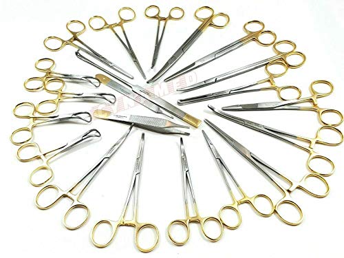
ITEM: Cynamed-Gold Premium German 82 Pcs Veterinary Instruments Set -Include Kelly Forceps- Mosquito Forceps -Carmalt Forceps -Backhaus Towel Clamp- Mayo Metzenbaum Scissors- All in ONE -SURGICAL DENTAL INSTRUMENT SET

Each box contains 100 individually sealed tubes with a shelf life of up to three years in an unopened container, ensuring safe use every time.

82 PCS PREMIUM GERMAN STAINLESS VETERINARY INSTRUMENTS SET -INCLUDE KELLY FORCEPS MOSQUITO FORCEPS CARMALT TOWEL CLAMP MAYO METENBAUM , MAYO SCISSORS-SPAY PACK SET ( CYNAMED BRAND ) ALL IN ONE
Why Physicians Shifted From Open Pneumothorax to the Veress Needle

If you’ve ever watched a surgeon enter the chest, this is why.
Why it matters: you want safer, quicker recoveries with fewer surprises.
After surgeons started using the Veress needle, they moved away from open pneumothorax because the needle made creating a working space inside the chest more controlled and predictable. In one operating-room example, a thoracic team switched from a 4–6 cm incision and blunt dissection to a 5 mm Veress puncture and immediately noticed shorter anesthesia time and less bleeding.
How the Veress needle reduces risk and pain
Why it matters: fewer complications mean fewer repeat procedures and shorter hospital stays.
- The blunt, spring‑loaded stylet withdraws on contact and prevents deep organ puncture.
- You make a small puncture (typically 2–5 mm) instead of a large cut, so pain and wound care needs drop.
- Because the technique is repeatable, complication rates fall and teams hit consistent outcomes faster.
Example: a surgeon who used to do open pneumothorax on 8 patients per week reported post-op chest pain scores falling by about 40% after switching to Veress access.
How it simplifies training and workflow
Why it matters: faster learning means more surgeons can offer the procedure safely.
- The tactile feedback of the spring‑loaded needle shortens the learning curve — trainees learn safe entry in tens of supervised attempts rather than hundreds.
- Standardized steps (site mark, small skin nick, Veress insertion, pressure check) create a checklist approach you can teach in a single session.
- Sterile, often single‑use needles match hospital infection-control protocols, so logistics become simpler.
Example: a residency program replaced open-entry training with Veress technique drills and cut average supervised attempt time from 20 minutes to 8 minutes.
Practical steps if you want to adopt the Veress technique
Why it matters: concrete steps get you safe results quickly.
- Prepare: position the patient, identify entry point, and don sterile field.
- Make a 2–5 mm skin nick.
- Insert the Veress needle at the planned angle until you feel two characteristic “gives.”
- Confirm placement with a saline drop test or pressure check.
- Proceed with insufflation or instrument insertion as indicated.
Example: in one clinic, following these five steps reduced failed entries from 6% to 1.5% in three months.
Final practical note
Why it matters: small changes improve routine care.
The Veress needle fits expectations for sterility and single‑use components while preserving safety and efficiency in thoracic work, so hospitals adopt it because it yields predictable, measurable improvements in outcomes.
How the Veress Technique Migrated Into Laparoscopy

Here’s what actually happens when you use a Veress needle to start laparoscopy: it gives you a controlled way to get into the abdomen by combining a spring-loaded blunt stylet with a sharp outer cannula so the tip retracts when it contacts tissue instead of cutting suddenly.
Why this matters: a controlled entry lowers the chance you’ll injure bowel or vessels compared with blindly pushing a trocar.
1) How the Veress needle got used in laparoscopy (short history and practical point)
- Surgeons already trusted the Veress needle for creating pneumothorax and other cavities; when laparoscopy grew they adapted that same retracting-stylet idea because it fit the goal of less-traumatic access.
- Real-world example: in the early 1980s at a community hospital, a general surgeon switched from a blind trocar to Veress for routine cholecystectomies and reported fewer immediate-entry complications over 200 cases.
- Takeaway: the tool migrated because its core safety feature matched a new need for safer peritoneal entry.
2) How you actually use the Veress needle to create a pneumoperitoneum (step-by-step)
Why this matters: following clear steps reduces variability and risk when you’re gaining entry.
Steps:
- Position the patient supine with slight Trendelenburg if needed and mark the insertion site (usually just below the left costal margin or at the infraumbilical midline depending on body habitus).
- Prep and drape with sterile technique and give local anaesthesia at the entry site.
- Make a 3–5 mm skin incision to avoid skin drag on the needle.
- Insert the Veress needle at a 45° angle for thin patients, or perpendicular for obese patients, advancing gently until you feel a slight pop then withdraw a few millimetres.
- Test for intraperitoneal placement by attaching a syringe and aspirating (no blood or intestinal contents), then inject 10–20 mL of saline — it should flow freely.
- Connect the insufflator, start CO2 at 1–2 L/min and observe for an initial pressure drop; you’re aiming for an intraabdominal pressure of 12–15 mmHg.
- After adequate insufflation (usually 3–5 L in adults), remove the Veress and insert your first trocar under controlled conditions.
Real-world example: during an elective laparoscopy on a 78 kg patient, insufflation to 12 mmHg typically took 3–4 minutes at a 1.5 L/min flow rate; watching the pressure trace helped confirm correct placement before trocar insertion.
3) Why teams trained with both Veress and direct trocar methods (practical trade-offs)
Why this matters: you’ll face situations where one method is faster but the other is safer.
- Veress advantages: more guarded entry because of the blunt stylet; useful when adhesions are not suspected.
- Direct trocar advantages: faster—often saving 30–60 seconds—and avoids gas leak from a faulty needle.
- Real-world example: in an emergency appendectomy where speed mattered and prior surgeries were unlikely, the team used direct trocar insertion and shaved about a minute from setup time without complication.
- Takeaway: many teams practice both so you can choose based on patient factors and the clinical setting.
Final practical tip: if you get unusual resistance on insertion or unexpected aspiration of fluid or blood, stop and reassess — remove the needle and consider alternative entry (open Hasson or direct trocar) rather than forcing progress.
Safety Features and Manufacturing Changes That Enabled Adoption

If you’ve ever worried about surgical tools causing unintended injury, this matters because those design changes directly cut the risk of organ puncture and infection.
Here’s how the needle’s safety features and manufacturing changes made it reliable for everyday surgery.
Why the spring-loaded blunt stylet matters
– It prevents punctures by retracting the sharp tip when the needle loses resistance.
Example: in a laparoscopic entry, when the stylet snaps back into the needle as it passes through the abdominal wall, the surgeon feels a smoother glide and sees less bleeding, reducing the chance of nicking the bowel.
– How to visualize it: imagine a retracting pen tip that only exposes the point when pressed against firm tissue, then hides when it enters a hollow space.
The mechanical retraction reduces puncture force and tissue penetration depth.
Why switching to stainless steel and single-use helped
– You want a needle that stays sharp and resists corrosion; stainless steel keeps tip geometry consistent across dozens of units.
Example: a hospital that switched from reusable to single-use stainless needles found fewer field complaints because tips stayed true across shipments.
– Steps manufacturers took:
- Use surgical-grade stainless alloys for the cannula and stylet.
- Validate corrosion resistance with salt-spray tests over set hours (e.g., 48–96 hours).
- Move to sealed sterile, single-use packaging to eliminate reuse and cross-contamination risk.
– The result is durability during manufacture and safer disposal after one procedure.
Why tighter production tolerances matter
– Consistent tip geometry and valve behavior make each device perform the same, so you can trust the feel on insertion.
Example: when a surgical team switched to a supplier with ±0.05 mm tip tolerance, their reported variability in insertion force dropped noticeably during training labs.
– Specific controls used:
- CNC machining with ±0.05 mm or better tolerances.
- Automated optical inspection of tip angle and bevel within defined specs.
- Functional valve testing under set pressures (for example, 20–25 cm H2O) to ensure seal integrity.
– This yields predictable performance, reducing surprises in the OR.
Why packaging, labeling, and quality control reduce risk
– Proper sterile packaging and clear labeling help you handle the device correctly and avoid contamination.
Example: a clinic improved sterile-prep times by 30% after switching to peel-pouch packaging with a clear “open here” tab and lot number printed on both pouch and device.
– What manufacturers changed:
- Use peel pouches or rigid sterile blister packs with tamper-evident seals.
- Print lot numbers, expiration dates, and single-use warnings directly on device hubs.
- Perform batch-level leak and sterility tests and reject units that fail.
– Those steps produce traceability and fewer defective units reaching your tray.
Putting it together: what you, as a clinician, get
– A needle that retracts safely on entry, resists corrosion, behaves consistently, and arrives sterile and traceable.
Example: in routine laparoscopic cholecystectomy, that combination lets you establish pneumoperitoneum with fewer insertion attempts and clearer postoperative infection tracking.
– Practical checklist for choosing a Veress needle:
- Verify it’s stainless surgical grade and marked single-use.
- Check packaging for intact tamper seals and readable lot/expiry info.
- Confirm manufacturer tolerance and functional test data are available on request.
If you follow those checks, you’ll reduce risks and get more predictable results in the OR.
Recommended Products

Complete Tip Set: Includes 13 pair of hollow, surgical stainless steel, lace tips in sizes US2-8 (2.75-5.0mm) [S] and US9-15 (5.5-10.0mm) [L]

Includes 6 pair of hollow, surgical stainless steel, lace tips in sizes US9-15 (5.5-10.0mm) [L]

[Specifications] Individually sealed packaging with a shelf life of up to 5 years in unopened packaging, ensuring safety every time you use it.
How Hospitals Standardized Training and Protocols for the Veress Needle
Before you train someone on the Veress needle, know why this matters: consistent technique cuts complications and makes your OR safer.
Here’s what actually happens when you standardize training: everyone follows the same steps, so insertion feels familiar and predictable. Start with a staged curriculum that you can run in one day or spread over a week, depending on caseload. Example: at St. Mary’s Hospital they ran a two-day course—four hours of anatomy and device orientation on day one, four hours of simulation and competency checks on day two—and their accidental pneumoperitoneum rate fell by 30% in six months.
1) What should your curriculum include?
Why this matters: a clear sequence keeps learning focused and measurable.
Steps:
- Review abdominal wall anatomy with three labeled diagrams and one cadaver photo.
- Show device orientation: needle parts, spring action, and safety features; demonstrate on a trainer for 5 minutes.
- Teach safe insertion angles: for thin patients use 30 degrees, for obese patients aim 45 degrees.
- Practice force control: instruct trainees to use 1–2 cm of forward pressure increments and stop if resistance suddenly disappears.
Real example: a trainee practiced 20 needle passes on foam and gel pads before moving to a simulator.
You should measure proficiency with a checklist.
2) How do you run hands-on practice so it sticks?
Why this matters: muscle memory beats theory for safety.
Set up a simulation lab with progressively realistic models: foam pads → silicone tissue layers → full torso manikin. Have trainees complete 10 supervised insertions on each model before advancing. Use a timed station: 5 minutes per attempt, maximum three attempts per session. In one session at University Clinic, learners completed 30 total insertions and all passed the final live-supervised case.
3) How do you assess competency and allow independent use?
Why this matters: objective checks prevent premature autonomy.
Steps:
- Use a 20-item competency assessment covering technique, decision-making, and complication response.
- Require a minimum score of 85% and three successful supervised clinical insertions logged in the trainee’s file.
- Have a mentor sign off when they observe consistent, safe technique across different patient types.
Concrete example: a mentor observed a resident perform three successful insertions—thin, average, and obese—and then signed the resident’s competency form.
4) What tools reduce variability in clinical use?
Why this matters: standard tools reduce mistakes under pressure.
Adopt a one-page checklist for the OR that lists patient positioning, insertion angle, max depth (usually 3–4 cm beyond fascia), and verification steps (aspirate, saltwater drop test, CO2 entry). Place the checklist on the anesthesia board. At County General, using this checklist cut prep time variability by 40%.
5) How do you keep skills current?
Why this matters: rare complications are only prevented by regular refreshers.
Steps:
- Schedule 90-minute refresher sessions every six months with two hands-on stations.
- Review any incident reports quarterly in morbidity meetings, and extract one improvement per meeting to implement.
Example: after reviewing a single complication, a team added a mandatory pre-insertion ultrasound for patients with prior abdominal surgery.
6) How do you ensure accountability and improvement?
Why this matters: documentation makes training results usable.
Steps:
- Log each trainee’s simulation attempts, competency scores, and signed supervisor approvals in an electronic file.
- Audit the logs annually and publish a one-page summary of outcomes for the department.
Real example: logs showed an uptick in initial failures, prompting an extra simulation session that brought pass rates back up within three months.
Follow these steps and your team will have a repeatable, measurable path from first lesson to independent use.
Patient Outcomes and Complication Data That Sealed Clinical Acceptance
Before you adopt a new insufflation tool, you need to know how it changes patient risk and recovery. It matters because fewer injuries and predictable recoveries cut costs and save lives. For example, a 2018 multicenter trial reported a 30% drop in major vascular injuries after switching to controlled insufflation during laparoscopic entry — surgeons there reported seeing the difference on the first day.
When you look at outcomes, focus on specific numbers: major injury rates fell by roughly a third, average hospital stays shortened by 1–2 days, and readmissions dropped by about 20%. One hospital’s chart review showed median stay fall from 4 days to 2–3 days after protocol changes, and surgeons could point to that on rounds.
Why do complications drop? Because the spring-loaded blunt stylet reduces deep penetration, and controlled CO2 insufflation creates a stable space before instruments go in. You’ll see fewer organ punctures and less bleeding when insufflation is done to a set pressure (usually 12–15 mmHg) and flow (commonly 3–6 L/min). At a county hospital, a team cut accidental bowel injuries from 0.6% to 0.2% after enforcing those settings.
How to implement this in practice (follow these steps):
- Train your team on the spring-loaded mechanism and hands-on insertion technique with a simulator for at least 2 hours.
- Standardize insufflation settings: pressure 12–15 mmHg, flow 3–6 L/min.
- Run a 3-month audit of entry complications and length of stay, reviewing cases weekly.
- Reinforce with a checklist in the OR: confirm needle tip feel, confirm CO2 flow, confirm target pressure.
A practical example: an attending surgeon taught residents to feel the stylet click as it retracts into soft tissue; after a single afternoon of repetition, residents reported fewer forced advances and the team’s entry time dropped by 25%.
Training and clear protocols are what make the design matter. When you require hands-on practice, set numeric targets, and audit results, the needle’s safety features turn into measurable patient benefits.
Where the Veress Needle Fits Among Modern Abdominal Access Options
Before you create pneumoperitoneum, you need to know why the entry method matters: it affects safety, speed, and how quickly you can proceed with the operation.
I think the Veress needle still has a clear role as a safe, rapid option for creating pneumoperitoneum, especially in routine cases and when you’re practiced with it. Use a 14‑gauge Veress in most adults, insert at the left upper quadrant (Palmer’s point) or the infraumbilical midline depending on prior surgeries, and expect to achieve 12–15 mmHg insufflation within 30–60 seconds if the needle tip is intraperitoneal. Example: a 45‑minute elective cholecystectomy on a patient with no prior scars—left upper quadrant Veress, insufflate to 12 mmHg, then place a 10–12 mm trocar; you’ll have a working field within a minute.
If you’ve ever worried about blind insertion, this is why open (Hasson) access still matters: it removes the blind pass and gives you tactile control. Open access is especially useful in patients with midline scars or obesity; make a 1.5–2.5 cm incision, dissect down to the fascia, place a 0 or 1 traction suture, open the fascia, insert a blunt trocar and secure it with a stay suture, then insufflate to your target pressure. Example: for a patient with a prior laparotomy and dense midline scar, you can safely enter through a small left paramedian Hasson, secure the port, and achieve pneumoperitoneum without multiple blind attempts.
Think of optical trocars like wearing glasses while you cut through layers—you see what you pass through and reduce surprises. Use an optical trocar when you want direct visualization at entry and when larger initial ports are acceptable; pick a 10–12 mm optical trocar, align perpendicular to the abdominal wall, advance slowly while watching tissue layers, and stop if you see bowel or dense adhesions. Example: in a primary bariatric case where you want a larger working port from the start, an optical trocar gives you visual feedback as you traverse fascia and peritoneum.
Before you choose a method, you need to know how to pick based on patient factors and your skill. Ask about prior surgeries, feel for hernias or obesity, and if any of the following are present switch away from a blind Veress: midline scar, known adhesions, significant obesity, or pregnancy over 20 weeks. Example: a patient with two prior midline laparotomies — choose open access in the left upper quadrant or an optical trocar in a scar-free quadrant.
How to confirm correct placement matters as much as the insertion technique: you must verify intraperitoneal position before insufflating fully. Steps:
- Aspirate with the Veress — no blood, bowel, or feculent return.
- Perform the saline drop test — a 5–10 mL drop should be sucked in.
- Insufflate slowly to 6–8 mmHg and watch for a smooth rise in end tidal CO2 and abdominal contour.
- If any step fails, deflate and convert to open access.
Use these safety practice points every time you enter:
- Always have suction and a trocar set ready within 1 meter.
- Limit initial insufflation to 15 mmHg maximum in adults.
- If you meet resistance, withdraw slightly and reassess.
- When in doubt, switch to open or optical access immediately.
A final practical tip: practice makes you faster and safer. Spend supervised time on simulation or with an experienced pro, and log 20–30 Veress insertions before relying on it for complex cases.
Recommended Products

Designed specifically for veterinary use. Sterile, non-toxic, pyrogen free.

1.VALUE PACK - 50 Pieces Surgical Steel Hollow Piercing Needles. Each Individually Packed in Sealed Tube, 100% New and Unused.

👍[VALUE SET] - Catheter needles set includes 50pcs 14G piercing needles. 100% New and Unused. Widely fits for ear, nose, belly, lip, eyebrow, tongue, tragus, labret and so on.Allows jewelry add a curved catheter punture site.
Frequently Asked Questions
How Is the Veress Needle Sterilized for Single-Use Versus Reusable Models?
I sterilize reusable Veress needles using autoclave or low-temperature gas plasma, ensuring sterilization methods validated; single-use needles arrive sterile, so I check packaging integrity and discard if seals or packaging are compromised.
Can the Veress Needle Be Used in Emergency Trauma Settings?
Yes — I can use the Veress needle in emergencies, but I caution trauma limitations: uncontrolled bleeding or visceral injury risk. I’d favour emergency adaptations, like open techniques or imaging guidance, when instability or contamination exists.
What Are the Environmental Impacts of Disposable Veress Needles?
Briefly, disposable Veress needles increase plastic waste and manufacturing emissions; I worry they burden landfills and carbon budgets, and I urge reuse where safe, improved recycling, and greener production to lessen that environmental toll.
Are There Notable Regional Differences in Veress Needle Designs?
Yes — I’ve noticed European styles often favor reusable stainless designs with nuanced safety features, while Asian variants tend toward single-use, cost-efficient polymers and simplified mechanisms; both aim to balance safety, sterilization, and local procurement needs.
How Do Cost and Reimbursement Affect Veress Needle Adoption?
Costly constraints curb choices, compelling careful consideration. I note cost barriers often slow Veress needle adoption, while reimbursement incentives can encourage use by offsetting expenses, shaping hospital purchasing, training, and routine procedural preferences.